AI in Digital Pathology: Telling which is which in adenocarcinoma
Written by Ilgar Guseinov and Bryan Zafra on 12.01.2023
Artificial intelligence (AI) is revolutionizing the field of healthcare, and digital pathology is no exception. With the ability to analyze vast amounts of data, AI has the potential to significantly improve the accuracy and efficiency of pathology diagnoses. In this blog post, we will explore med4PAN’s current use case of AI in digital pathology. We will also explore the ethical considerations and challenges facing its implementation.
One of the main applications of AI in digital pathology is image analysis. By using machine learning algorithms, AI can analyze microscopic images of tissue samples and identify patterns that may indicate the presence of disease. This can be particularly useful in the diagnosis of cancer, where the identification of specific cell patterns is crucial for accurate diagnosis.
One of Med4PAN’s use case is to be able to differentiate adenocarcinoma from different anatomical sites e.g. breast, colon, esophagus, lung, and prostate. Pathology images were processed and standardised to be 256 x 256 pixels. A base template AI model from Google Tensorflow Hub called efficientnet_v2 is used in training med4PAN’s model for digital pathology. Overall, there are 207 million parameters from these pathology images of which 6,405 parameters are trainable to be incorporated to the AI model. The resulting AI model training process has an accuracy of 99.64 percent.

Figure 1. Training and validation loss function (above) and accuracy (below) of the AI pathology model.
The model also has good predictions in differentiating adenocarcinoma from different anatomical sites. For example, the table below showed that the images number 1 to 3 have high prediction score of more than 99% while image number 4 have a prediction score of 95%. However, for the image number 0, the AI model was unable to make high predictions and confuses it between adenocarcinoma of the lung or the esophagus.
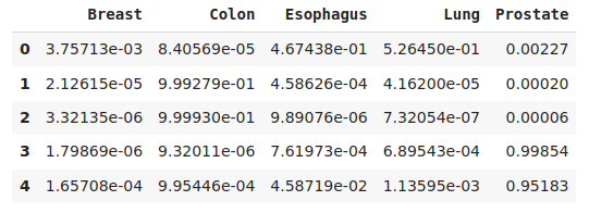

Table 1. Sample predictions of the AI model showing the first 5 images of the dataset and their corresponding prediction scores for each type of adenocarcinoma.
While the potential benefits of AI in digital pathology are significant, legal considerations must also be taken into account. Privacy concerns are of particular importance because pathology images often contain sensitive personal information. For example, the DICOM format may contain some personal information such as name, date of birth, etc. And here it is especially important to comply with GDPR standards. In addition, there is the possibility of bias in the AI system, especially if the training data is not representative of the population to whom it is used for diagnosis.
Another problem standing in the way of implementing AI in digital pathology is the lack of standardization. Currently, there is no standard method for creating, annotating, or sharing digital pathology images, which can make it difficult to create AI systems that can be used across institutions. In addition, there is a lack of data to train AI systems, which can make it difficult to achieve high accuracy. A whole slide scanner will be used to collect the data that will be used to train the model and connected to the 5G network, which is currently being installed on the ECRI campus.
Despite some challenges, the future possibilities of AI in digital pathology are very interesting and with the development of 5G networks will be much more accessible, but we will talk about that in our next posts!